
By Gwen Rockwood
Most mothers and expectant mothers know the intense mix of emotions you feel while lying on the exam table, waiting for your ultrasound to begin. Part of you is so excited you can hardly wait, and the other part of you is so scared you’re holding your breath. Your mind races with twenty questions at once: Is it a boy or a girl? Will we see 10 fingers and toes? Is she healthy? What if something is wrong?
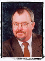
With the anxiety and excitement swirling together, it’s good to have someone calm behind the scope of the ultrasound machine – someone who’s trained to give your unborn baby as thorough a check-up as ultrasound technology will allow. That’s where Dr. Lindley Diacon comes in. He and his team of sonographers at Mercy Health System have been giving parents a glimpse into the womb to see how their baby is developing. Though he downplays his national renown, Dr. Diacon is actively shaping the way ultrasound technology is used nationwide. He sits on the board of the Practice Accreditation Council of the American Institute of Ultrasound in Medicine and is one of the physicians writing the exam all ultrasound technicians must pass to be certified.
How does it work?
Ultrasound works by using sonar waves. These sound waves are sent into the body and reflected back, providing a picture. In recent years, we’ve begun to hear references to different “levels” of ultrasound exams, like Level 1 or Level 2. Dr. Diacon said the difference between a Level 1 and Level 2 exam is pretty simple. Level 1 is a basic exam, and Level 2 is a more detailed exam that screens for problems more difficult to spot.
Patients with extremely high risk pregnancies often see a perinatologist – a physician with extensive training in high risk obstetrics as well as ultrasound technology. (Currently there are no perinatologists practicing in Northwest Arkansas, although there are some in Little Rock, Joplin and Springfield.)
To ensure that your ultrasound is being done by someone qualified, you can ask if the person is a registered sonographer with the American Registry of Diagnostic Sonography.
3D and 4D Ultrasounds
Advancing technology in ultrasound equipment, such as 3-D and 4-D ultrasound, has enhanced the experience and provides some great pictures for the baby book. Mercy Health System recently bought a new 4-D machine that gives highly detailed photos of babies in utero. “You can really see the details of the face, so much so that you can sometimes see family resemblance,” Diacon said. “It’s really fun.”
But if you don’t have access to a 4-D ultrasound, Dr. Diacon said you shouldn’t worry that your exam is not as accurate. A basic exam on a machine without 3-D capability is still an excellent tool for catching problems early. “From a diagnosis standpoint, you’re not really missing anything if you can’t get a 4-D ultrasound,” Diacon said.
Ultrasound “Parlors”
Ten years ago ultrasound parlors didn’t even exist, but today it’s not uncommon to see a pregnant woman walk into a small shop in a strip mall and come out carrying photos of her unborn child. Dr. Diacon has reservations about this use of ultrasound and said the FDA has put out some strong statements about these types of places.
“You need to understand that these ultrasound parlors are not doing a diagnostic exam,” Diacon said. “These types of ultrasounds sometimes give parents a false sense of security.”
Medical advancements
The latest improvements in ultrasound technology mean good news for parents. Dr. Diacon said prenatal ultrasounds are getting better at detecting fetal heart defects. And early ultrasounds done between 11 and 14 weeks gestation are now better at diagnosing chromosome problems like Down Syndrome. (Ask your sonographer about the six “soft markers” for Down Syndrome.)
A Day In The Life
Believe it or not, Dr. Diacon does up to 30 ultrasounds a day. He said he usually starts his day doing ultrasounds on gall bladders, livers and abdomens and then moves on to vascular exams and OB patients. But it’s obvious when talking with him that expectant parents are a special part of his job.
“It’s great because 98 percent of the time, things are fine with the baby and I love talking to the patients. I get a kick out of watching them watch their baby,” Dr. Diacon said. “And when things are not fine, I can be there to share their sorrow and reassure the mother that it’s not her fault. That’s such an important thing for a woman to know.”
Sneak Peek at a Miracle
 A final question for Dr. Diacon: “What was your most memorable ultrasound and why?” He answered with this story:
A patient had a certain genetic condition that has a 50 percent chance of being passed to her children. The condition is so severe that any child born with it dies in infancy. The woman and her husband had already suffered through the loss of two babies but had not given up on the dream of having children. Dr. Diacon performed the ultrasound during their third pregnancy, and, after a long, tense examination, told the couple that this baby did not have the genetic disorder. This baby would live.
“I’ll never forget that moment,” Diacon said. “They were so overcome with joy and relief. I really bonded with them and, several months later, I even had the chance to be there during that baby’s birth.”
Dr. Lin Diacon is the head of the Central Imaging department for Mercy Medical Clinic. (Click here to read more about Dr. Diacon’s work with bone density scans.) For more information on ultrasound technology, visit the Fetal Medicine Foundation website. http://www.fetalmedicine.com/